Conquering breast cancer with a Trojan Horse
The antibodies shown here are produced inside cancer cells, and eat up a tumour from the inside. This battle of the cells takes place in iridescent colours.

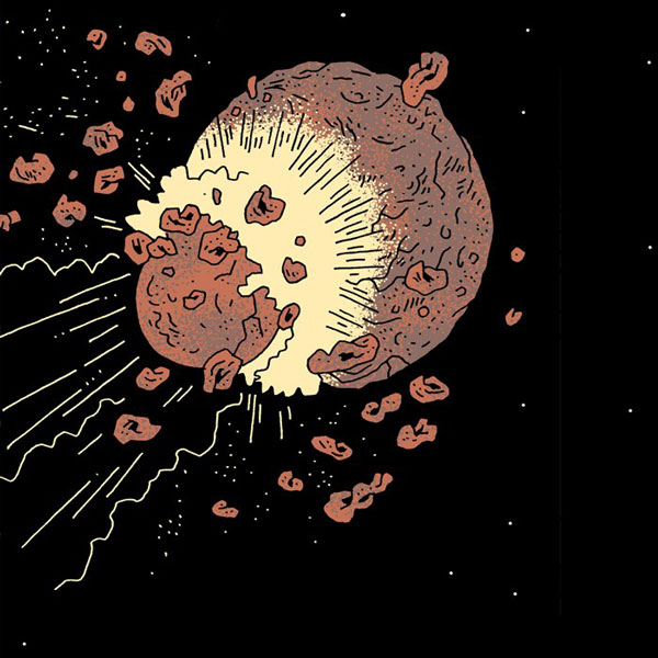
“What looks beautiful is actually the cancer cells; the black sections are where the therapy was successful. That’s paradoxical, which is also why I find this image attractive”, says the biochemist Sheena Smith. | Image: Sheena Smith, with assistance from Branko Simic and Rajib Schubert
A battle is taking place here. The attackers are still in the middle of their fight in the green spots; and where they’ve won, the result is blackness. “We like to use the analogy of the Trojan Horse”, explains the biochemist Sheena Smith. “We use a virus that smuggles genetic information into breast cancer cells. This information enables the production of an antibody that recognises tumour cells and kills them. It’s as if the cancer cells are being eaten up from the inside”.
This experimental gene therapy is being developed by Smith and her colleagues at the Plückthun Lab at the University of Zurich. By producing antibodies directly in the cancer cells themselves, they’re striking out in a new direction. This is also where Smith gets her sense of motivation: “We’re looking way over the garden fence of traditional strategies”.
The battle taking place on our microscopic photo was in the breast of a mouse, and it shows a cross-section of tissue. When the artificially developed virus infects the cell, the green areas occur. The blue dots are the nuclei of the tumour cells, while the red stripes are the blood vessels that nourish them. The cells that have died off remain completely black. “So what looks beautiful on the image is actually the cancer cells; the black sections are where the therapy was successful. That’s paradoxical, which is also why I find this image attractive”, says Smith. The image was captured in an early phase of their series of experiments, which is another reason Smith likes it so much: “It was really exciting to be able to observe how the therapy functioned for the very first time”.
In order to heighten the aesthetic impact of the image when it was submitted to the SNSF photo competition, the researchers intensified the way the colours glow. Smith explains how: “We took the fluorescent signals of the different parts of the cells and transferred them into colours that look good on a screen. But they also correspond largely to the general colours that we use to visualise things in our research”. This iridescent snapshot has rather fantastical associations for Smith: “It could almost be the night sky in a science-fiction film”.